 |
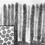
Microvilli of normal enterocyte cut
Microvilli of normal enterocyte cut longitudinally and transversally (inset). Electron micrograph, scale = 0.2 mm.
|
|---|
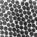 |
Microvilli of normal enterocyte cut
Microvilli of normal enterocyte cut transversally. Electron micrograph.
|
|---|
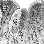 |
Normal epithelium of the small intestine
Normal epithelium of the small intestine: E - enterocyte, G - goblet cell. Low magnification electron micrograph, scale = 2 mm.
|
|---|
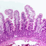 |
Normal light micrograph of small intestine
Normal light micrograph of small intestine - jejunum. Mucosa shows tall fingerlike villi and short crypts. Villi are covered predominantly by the absorptive cells(cells with brush border) but also contain several goblet cells. Muscularis mucosae can be seen on the bottom of the sample. HE
|
|---|